

The female reproductive structures of a flowering plant develop within the ovary. The ovary contains one or more ovules, and each ovule provides the environment in which the female gametophyte develops.
To understand how the female gametophyte is formed, we first need to look at the structure of an anatropous ovule.
Structure of an Anatropous Ovule
The ovule is attached to the placenta inside the ovary by a stalk called the funicle. In most flowering plants, the ovule is inverted and is known as an anatropous ovule.
A typical anatropous ovule has several important parts.
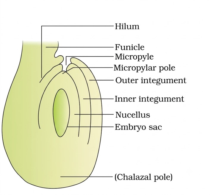
The funicle is the stalk that connects the ovule to the placenta. The point where the body of the ovule is attached to the funicle is called the hilum.
The ovule is surrounded by one or two protective coverings called integuments. These integuments do not completely enclose the ovule. They leave a small opening called the micropyle.
Opposite the micropilar end, is the chalaza, representing the basal part of the ovule.
Inside the integuments is a mass of tissue called the nucellus. The nucellus contains the developing female gametophyte, or embryo sac.
The main parts of an anatropous ovule include the funicle, hilum, integuments, micropyle, nucellus, chalaza and embryo sac.
Formation of the Female Gametophyte
The development of the female gametophyte begins with a specialised cell within the nucellus called the megaspore mother cell.
The megaspore mother cell undergoes meiosis and produces four haploid megaspores. This process is called megasporogenesis. Usually, only one of the four megaspores remains functional, while the other three degenerate. The single functional megaspore then undergoes a series of mitotic divisions and develops into the female gametophyte, also called the embryo sac.
This type of embryo sac development, in which the embryo sac develops from a single functional megaspore, is called monosporic development.
The basic sequence is:
Others Degenerate
7 Cells, 8 Nuclei
Development of the Embryo Sac
The functional megaspore does not immediately become a mature embryo sac. It first undergoes mitotic divisions. The nucleus of the functional megaspore divides once to form two nuclei. These move towards opposite poles of the developing embryo sac.
Further mitotic divisions produce four nuclei and then eight nuclei. The eight nuclei become organised within the embryo sac to form the mature female gametophyte.
A typical mature embryo sac is 7-celled and 8-nucleate.
Organisation of the Mature Embryo Sac
The mature embryo sac has three important regions.
At the micropylar end, there is the egg apparatus, consisting of three cells:
- One egg cell
- Two synergids
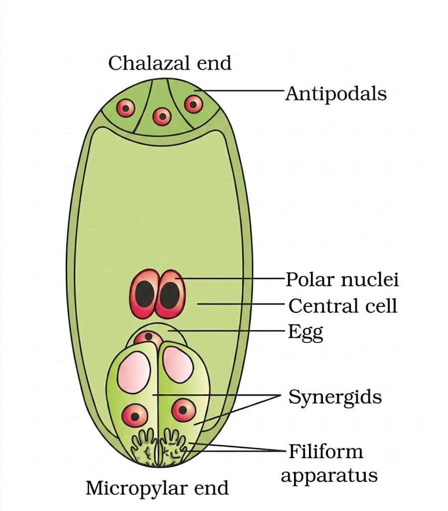
The synergids possess special structures called the filiform apparatus, which help guide the pollen tube towards the egg apparatus.
At the opposite, chalazal end, there are three antipodal cells.
The remaining two nuclei, called the polar nuclei, are located in the centre of the embryo sac. They may fuse to form the secondary nucleus before fertilisation.
Therefore, the mature embryo sac contains:
Cells: 3 antipodals + 1 central cell + 2 synergids + 1 egg = 7 cells
Nucleus : 3 in antipodals + 2 polar nuclei + 2 in synergids + 1 egg = 8 nucleus
The two polar nuclei belong to the same central cell, which is why the embryo sac has seven cells but eight nuclei.
From Megaspore to Mature Embryo Sac
The entire development can be followed as a simple sequence:
Megaspore mother cell → Meiosis → Four haploid megaspores → One functional megaspore → Three mitotic divisions → Eight nuclei → Organisation of nuclei into cells → Mature 7-celled, 8-nucleate embryo sac
This mature embryo sac is now ready to participate in the events of fertilisation.
Important Points to Remember
- The ovule is the megasporangium of a flowering plant.
- The most common type of ovule found in angiosperms is the anatropous ovule.
- The ovule is attached to the placenta by the funicle.
- The point of attachment of the funicle to the ovule is the hilum.
- The small opening left by the integuments is the micropyle.
- The tissue inside the integuments is the nucellus.
- The megaspore mother cell undergoes meiosis to produce four haploid megaspores.
- The formation of megaspores from the megaspore mother cell is called megasporogenesis.
- The functional megaspore undergoes three mitotic divisions to produce eight nuclei.
- The mature embryo sac is typically 7-celled and 8-nucleate.
- The egg apparatus consists of one egg cell and two synergids.
- The chalazal end contains three antipodal cells.
- The two polar nuclei are located in the central cell.
FAQs
An anatropous ovule is an inverted ovule in which the body of the ovule becomes oriented close to the funicle. It is the most common type of ovule found in flowering plants.
The main parts are the funicle, hilum, integuments, micropyle, nucellus, chalaza, and embryo sac.
Megasporogenesis is the formation of haploid megaspores from a megaspore mother cell through meiosis. Four megaspores are usually formed, but only one remains functional.
The megaspore mother cell undergoes meiosis and produces four haploid megaspores. Usually, one megaspore remains functional. It undergoes three mitotic divisions to produce eight nuclei. These nuclei organise into a mature 7-celled and 8-nucleate embryo sac. This is the female gametophyte.
A mature embryo sac has seven cells but eight nuclei. It contains three antipodals, two synergids, one egg cell, and one central cell. The central cell contains two polar nuclei, giving a total of eight nuclei.